
What is Gut Stasis?
As most rabbit owners will be only too willing to testify, pet rabbits can be very susceptible to health problems, many of which are difficult to diagnose and costly to treat. Since the start of CottonTails rabbit and guinea pig rescue in 1993, the primary health issue that has given us the most cause for concern apart from dental disease has been the killer diseases gut stasis, bloat, and mucoid enteritis. Although these conditions are also referred to as ileus, gastrointestinal (GI) stasis and bloat, there are important distinctions between these conditions.
In most cases, it would appear that GI stasis/ileus develops slowly, usually several days before you suspect there is a problem. Still, if diagnosed early enough, the condition is often treatable. Bloat, however, happens suddenly and without warning, and the prognosis for survival is poor, although bloat can even develop as a complication of GI stasis (1). Ingestion of hair (fur ball) was initially thought to be a cause, but it has now been found that fur has gathered due to initial problems with gut motility and is not the causative factor (2).
Having said that, if a rabbit has had gut stasis issues before, it makes sense to groom out any excess fur if the rabbit is having an extremely heavy moult, as consuming excessive amounts of fur either from the rabbit itself or from a partner will not be helpful in such individuals and may be the tipping point for the development of another episode of stasis in addition to the problem predisposing the rabbit to this condition in the first place.
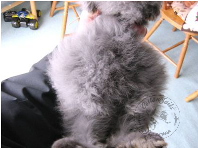
The photo below shows a young rabbit 15 weeks old that showed typical symptoms of bloat – he sadly died 8 hours later despite intensive treatment.
There have been several studies of mucoid enteritis carried out in rabbits, and there is general agreement that it is a severe condition which can affect whole herds of rabbits, not just individuals. One study (3) found that there was 60-70% mortality in young rabbits due to mucoid enteritis in an outbreak over two years. These figures are consistent with the findings at CottonTails.
Isolated incidents of mucoid enteropathy can often be attributed to stress of one sort or another, especially the sudden change of diet and environment of young rabbits, as well as inadequate diet, coccidiosis, and conditions causing the rabbit to stop eating, such as pain after surgery and dental disease. However, the outbreaks described by various authors and also experienced at CottonTails appear to be different and would indicate the involvement of an infectious element of some kind or some unknown “trigger” for the release of Clostridium botulinum toxin, the presence of which we have had confirmed at post mortem. Coccidiosis was not a contributing factor in those animals examined at CottonTails.
Symptoms and Possible Causes
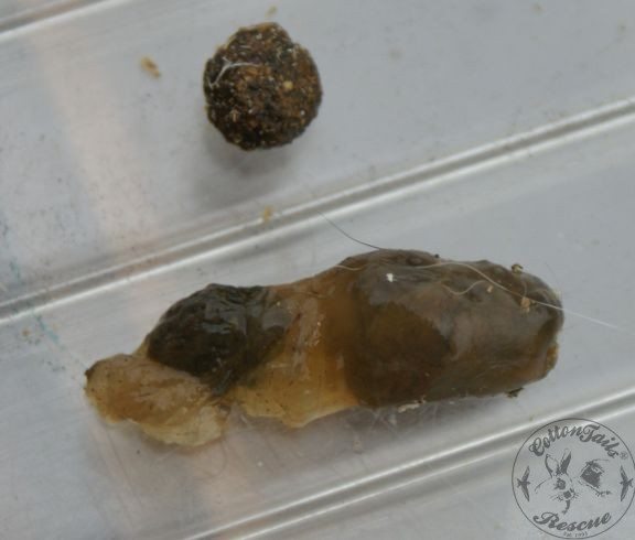
From personal observations, the onset of symptoms was usually very sudden, with loss of appetite, few or no droppings being passed, lying stretched out or huddled in a corner with apparent abdominal pain, and behaving in an abnormally quiet or withdrawn manner. A decrease in normal abdominal sounds or frequent gurgling was found, with subsequent development of palpable hard areas within the intestines often accompanied by the onset of bloat/and or diarrhoea and passing of mucus. The photo below shows a normal dropping beside a dropping encased in jelly-like mucus, indicating severe gut inflammation.
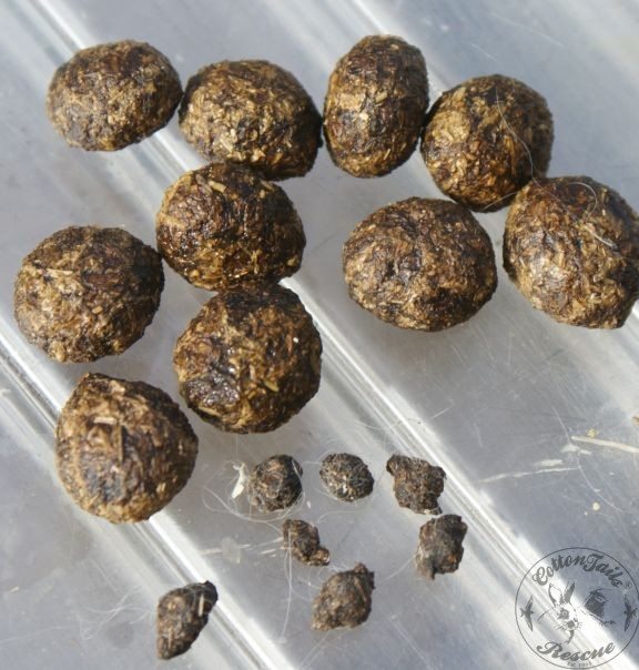
The photo below shows the difference between normal-sized droppings and those of a rabbit in the later stages of gut stasis just before he stopped passing droppings altogether. Note the small, hard and misshapen droppings, indicating that gut motility has slowed. The rabbit usually passed normal droppings, but the droppings got progressively smaller over a week.
The day immediately before the onset of symptoms, the rabbit would appear to eat and behave normally. The next day, it would be seriously ill and deeply distressed. However, the illness likely develops for some time before any obvious outward signs become apparent, making this condition very difficult to treat as often, by the time it is noticeable, it is then too late for the rabbit to be saved.
The first mucoid enteritis outbreak experienced at CottonTails occurred in 2002 when a group of 25 rabbits was brought in. They were part of a herd of over 100 rabbits of mixed sexes, breeds and ages, all previously running free in a garden. Most of the females were pregnant or had young litters with them. As a result, we had an exceptionally high number of young rabbits at the centre during this time. Several of the rabbits also had dental disease, and most were thin with wounds consistent with fighting. However, they all had good appetites and appeared fit and well.
Before the outbreak, no sudden changes had been made to diet or environment to explain the onset of the symptoms apart from when they arrived. The rabbits were all fed a vet-recommended measured diet of Burgess Excel, with fresh vegetables as appropriate for age and tolerance, and good quality hay and fresh water (from a drinking bottle) available at all times. Care was taken to avoid overcrowding, and litters were kept separate, usually with their mothers. Symptoms did not coincide with the timing of vaccinations or neutering, nor was there a pattern of infection based on hutch location as symptoms appeared in rabbits several hutches apart (in one case 150 feet away), the rabbits in between not being affected.
From the information gathered during this and another two outbreaks as well as random isolated cases, it would appear that the average duration of the disease from the point of first symptoms to either the start of recovery or death was 12 hours to 9 days, giving an average of 5 days. Of the total number of rabbits in the centre at the time, 20% were affected, most of which were rabbits under five months old or nursing mothers, with a mortality rate of up to 60%.
Very few adult rabbits became ill and had a much better recovery rate than the young rabbits, with 70% making a full recovery compared with 40% for those younger than five months. The incubation period varied from 6 days to 4 months, making quarantine impractical for future disease management.
Treatment Given
The affected rabbits were given intensive treatment: administration of Metacam for pain relief (plus Tagamet syrup in some cases to prevent possible inflammation of the stomach); Prepulsid to stimulate stomach emptying (not now available); Metaclopromide to stimulate gut motility (very effective but if a severe blockage is present, rupture of the stomach can occur); subcutaneous fluids to prevent dehydration (not effective in cases of shock, so some individuals received i.v. fluids); syringe feeding of Critical Care to those individuals willing to eat; Questran (Cholestyramine which absorbs enterotoxins (4)); subcutaneous administration of broad-spectrum antibiotic Baytril in some individuals to rule out infection; Kaogel in cases of diarrhoea; Vitamin B injection in some individuals in an attempt to stimulate appetite.
Gentle massage of the abdominal area was also carried out in those rabbits that would tolerate it. An incubator was used for individuals whose body temperature dropped significantly below normal (usually an indication that death was imminent). Personal observations indicated that recovery or death was not significantly influenced by medications, apart from the noticeable relief that Metacam provided.
By the time the third major outbreak occurred two years later, E. cuniculi, the protozoan parasite infecting the renal and nervous system of rabbits, had been officially recognised as a severe problem in the general pet rabbit population in the UK, possibly affecting at least 50% of pet rabbits (5). As a result, one of the affected litters that demonstrated the additional symptoms of hind leg weakness and white eye spots was tested for E. cuniculi and was indeed found to be positive.
With this in mind, it was decided to treat all rabbits with Panacur (orally, at a dose of 20mg/kg/day) to ensure that they were free of the parasite based on the suspicions that E. cuniculi could be an essential factor in turning what would ordinarily be an isolated occurrence of GI stasis into an infectious outbreak if only in respect that infected animals would have lower disease resistance. Panacur (fenbendazole, metabolised into the active form, oxfendazole) works by inhibiting an essential part of the feeding/infective apparatus of microsporidian parasites such as E. cuniculi (6).
Unfortunately, although a four-week course of Panacur has been shown to eliminate the parasite (given via their pelleted food) (7), there is no evidence to suggest that it can reverse neurological damage already done. However, if some of the symptoms are due to inflammation and the immune response, elimination of the parasites and anti-inflammatory drugs such as Rimadyl may be helpful.
The controversial study that indicated Panacur and its derivatives could cause bone marrow failure in a very small percentage of dogs, cats, birds, reptiles, and three unrelated rabbits (8) is not thought to be representative of the majority, and CottonTails will continue with our policy of administrating Panacur prophylactically, and there would appear to be more advantages than disadvantages in following current procedures.
However, Intervet has conducted a safety study to address the concerns of bone marrow problems, and initial studies have shown no evidence of any changes to haematological or biochemical parameters. They have launched a new product for E. cuniculi prophylaxis, especially for rabbits. This is in the form of a paste, and the recommended prophylactic regime will be a 9-day course 2-4 times yearly. It will also be recommended at high-risk times, such as when a rabbit is acquired, before mixing with new rabbits and before mating.
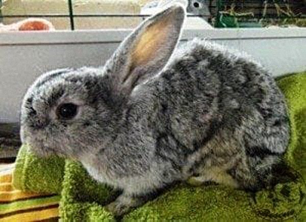
Based on personal communication with Intervet, the Panacur paste is ideal for rabbits over 1.25kg. Therefore, treating baby rabbits under this weight would still be best with 10% Panacur to achieve higher accuracy for the nine days required. The photo below shows a young rabbit struggling to survive gut stasis followed by diarrhoea, and despite all efforts, she lost her battle shortly after the photo was taken. She had lost a significant amount of weight.
Since the policy of administrating Panacur to all rabbits was initiated, CottonTails has had no further outbreaks of mucoid enteritis-type illness. There have been isolated individual cases, as one would expect in a rescue centre, where animals often arrive in poor condition and undergo changes in diet, environment, neutering and vaccination. Personal observations would indicate that either the Panacur itself or the elimination of the parasite has had some influence on this result.
In a personal communication with the small animal vet adviser for Intervet (the manufacturers of Panacur), it was suggested that the preventative effect we have witnessed may be due to the eradication of intestinal worms on the basis that a heavy infestation with Passalurus ambiguous in young rabbits can be a contributory factor to the enteritis complex of diseases that occur around weaning. As there has been no evidence of worm infestation in any of the affected rabbits, and none were found post-mortem, it seems unlikely that this is the case.
Other Factors
Another factor involved in the infectious element of mucoid enteritis could be bacterial infection leading to gastric ulceration, similar to Helicobacter pylori in humans (9). Personal observations showed that all rabbit fatalities examined from the mucoid enteritis outbreaks had erosion of the stomach lining, as seen by visible small circular areas. This was also found in a study that showed 7% of the rabbits examined had gastric ulcers (10). It is known that gastric ulceration in rabbits can develop from pain and fear due to catecholamine (epinephrine) release and reduced gut motility, such as repeated episodes of stasis (1).
As the broad-spectrum antibiotic Baytril was given by subcutaneous injection to some rabbits during the outbreaks and had no noticeable effect, it would appear that if a bacterial infection was involved, it may not be sensitive to this particular antibiotic. The possibility of a virus has been suggested in a study looking for viral agents in rabbits with enteropathy in Italy(11). The conclusion was that the cases of mucoid enteropathy-caecal impaction in several commercial rabbitries could not be explained by a “new” viral or bacterial agent with an enteric replication. Therefore, other factors not yet identified could be involved in causing or predisposing rabbits to this disease.
They couldn’t exclude, however, that the replication of viruses ordinarily present at a lower concentration might have a pathogenic action under certain conditions. There have been comparisons between mucoid enteropathy in rabbits and equine grass sickness in horses, whereby evidence showed that the origin of the disease in two particular cases involved soil-borne botulinum neurotoxin (12). This study showed for the first time that green grass blades can contain Clostridium botulinum toxin and put forward the hypothesis that equine grass sickness is a clinical form of botulism, a soil-borne disease.
The relevance of this finding to rabbits is in the large quantities of hay used routinely as an important food source. To add even more confusion to the situation, recent national and international research into gut-related problems in rabbits has identified a condition known as dysautonomia (13, 14, 15, 16), a severe disease affecting the autonomic nervous system. However, this is not thought to be the cause of the outbreaks at CottonTails. As the symptoms had some similarities, some of the rabbits suffering from mucoid enteritis were taken to Katherine Whitwell, a leading authority on dysautonomia in rabbits and hares, for her opinion. As the recorded symptoms and crude evidence did not indicate the presence of dysautonomia, further laboratory tests were not carried out.
Spotty Rabbit Syndrome Medication Update
There appears to be a genetic predisposition in certain breeds of rabbits to gut stasis issues, particularly in the English (Dalmation) type rabbit, but this does not mean that every spotty rabbit will have a problem! It is undoubtedly the case that once a rabbit has had a bout of gut stasis, it may be prone to further issues, possibly due to a narrowing of the gut due to the effects of the previous inflammation.
As yet, the cause of mucoid enteropathy-type outbreaks is still not known. Whether there could be some involvement of E. cuniculi can only be speculated, with infected rabbits being more vulnerable to disease. What is clear, however, is that research is urgently required if more rabbits are not to be lost to this killer disease.
Medication Update
My observations are that the following medications are the most helpful in cases of gut stasis: Metacam (usual dose for an average 2.5kg rabbit is 0.3-0.5ml once daily), Metoclopramide (gastric stimulant, dosage given orally is 0.5-1mg/kg every 6 hours) and Propulsid (Cisapride, hindgut stimulant) dosage 0.5mg/kg every 8 hours. If a blockage is suspected, it may be best not to use Propulsid unless the vet is agreeable. There is some evidence that Infacol (available from chemists in the high street) may be helpful if the problem is gas in the stomach or small intestine as it works by joining all the tiny gas bubbles into one bigger one, which is easier to pass along the digestive system.
References
- L. Seeman. Bloat in Rabbits.
- Susan Brown. Rabbit Hairballs: Fact or Fiction. Small Mammal Health Series. Veterinary Information Network Inc.
- Fur and Feather incorporating Rabbits, Health Club, no date, and comments by the Editor.
- Erica Deighton. Cholestyramine & Loperamide References.
- Emma Keeble. Study of E. cuniculi in rabbits.
- Dana Krempels. Hind Limb Paresis and Paralysis in Rabbits. Rabbit Health Central, Houserabbit Adoption, Rescue and Education (H.A.R.E.)
- Suter C. et al. Prevention and treatment of Encephalitozoon cuniculi infection in rabbits with fenbendazole. The Vet Record, April, 14, 2001 pp 478-480.
- Alexandra Logsdon, Zoo Corner Rabbit Rescue.
- Cesare Montecucco and Rino Rappuoli. Living Dangerously: How Helicobacter Pylori Survives in the Human Stomach. Nature Review, Molecular Cell Biology, MacMillan Magazines Ltd., Volume 2, June 2001.
- Hinton M. Gastric Ulceration in the Rabbit. J. Comp. Pathol. July: 90 (3) pp 475-481, 1980.
- Nieddu D. et al, Electron microscopy detection of viral agents in rabbits with enteropathy in Italy during the period 1982-1999. 7th World Rabbit Conference, 4-7 juillet 2000.
- Bohnel H. et al., Two cases of equine grass sickness with evidence for soil-borne origin involving Botulinum Neurotoxin. Journal of Veterinary Medicine B Vol 50 Issue 4 page 178, 2003.
- Whitwell K., Needham J. Vet Record 1996, 139 (13) 323-3
- Van der Hage M., Dorrestein G.M. Dysautonomia in rabbits (abstract) World Rabbit Science 1996 Vol 4(1), 10
- Kenneth Harkin, Media Relations and Marketing, October 2004.
- Feline Dysautonomia (Key-Gaskell Syndrome) study group.